Our city park has a very large eastern red cedar tree whose leaves were looking
a bit on the brown side. I also noticed other eastern red cedar trees in a
similar condition, so I took a sample branch from the park tree for analysis.
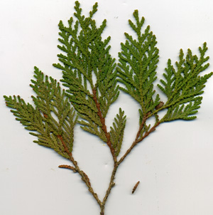
The branch is shown below, scanned at 300dpi, and sampled in late October.
Note: Commonly called Red Cedar, this tree is really a juniper, not a true cedar.
1
Note that the leaves on the left-most twig seem a bit browner, or a less healthy green.
On some trees, these twigs would turn totally brown and very easily break off.
One of the simplest analysis tasks is a microscopic look at the surface of a leaf.
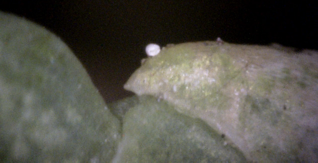
The following picture shows a leaf surface. Only one picture is shown, because, while
many were taken, they all showed little evidence of any disease.
2
The surface of all the leaves was generally the same as shown here - seemingly disease free.
Here, however, a small exception is shown - a single white canker spore.
Photos of the cross-section of a leaf are more difficult to obtain, but potentially show more.
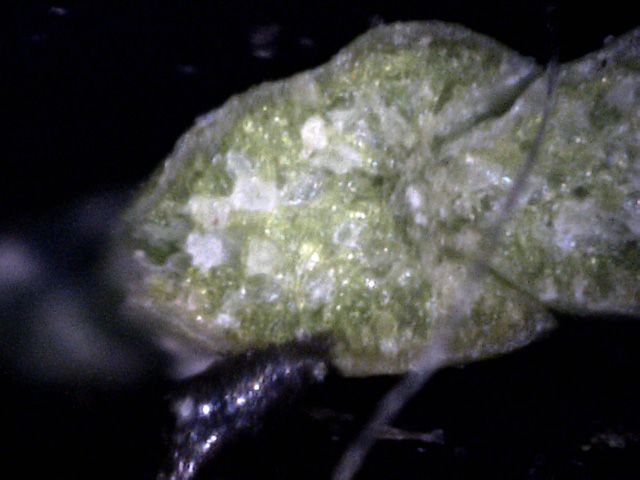
That's the case here, as these 400x photos of a tiny leaf show.
3
Note that there is lots of white canker present, but it's all buried within the leaf!
That's why it's presence is not noticed when looking at the surface of the leaf.
Hyphae are also present.
4
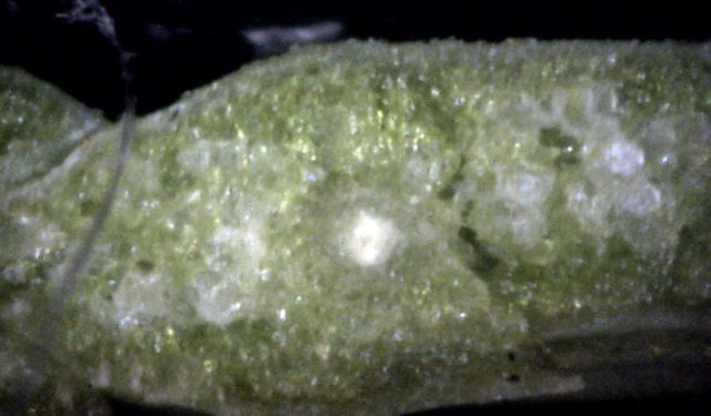
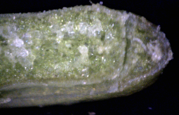
This is the middle of the leaf - again some canker, but it's buried deeply within the leaf.
The bright central spot is not white canker, but the leaf's main vein.
5
Once again, there is substantial white canker present, but it's all deep within the leaf.
There is also a blob at the right edge.
The examination of twig cross-sections usually gives better evidence of white canker than leaf cross-sections.
The following twig cross-sections were made with a 400x microscope. The twig was about 3/16 inch in diameter - a
convenient size for analysis.
 ←
←
←
←
←
←
6
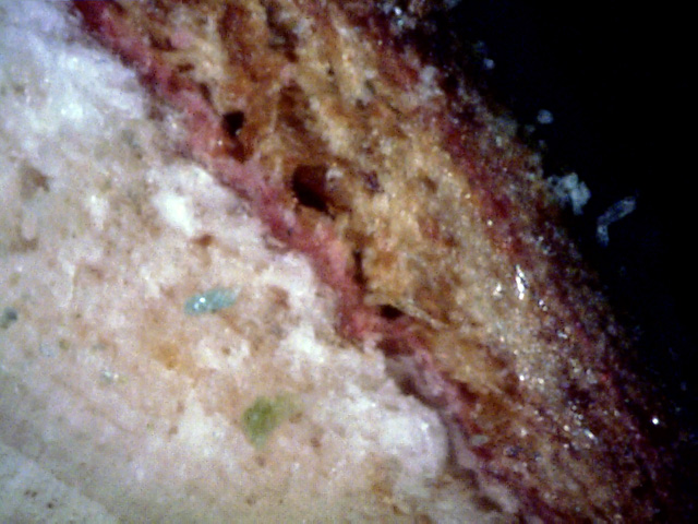
The outer edge of the phloem is almost all white canker (red arrow).
The cortex (blue arrow)seems pink for some reason.
But especially note the large amount of tan canker in the outer bark (yellow
arrow).
7
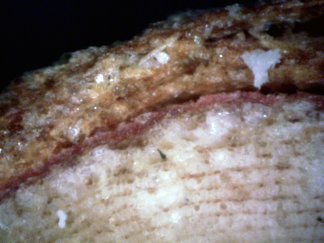
While the outer phloem here has quite a bit of white canker material present, it is more diffuse,
as in other conifers, rather than globular.
With the unaided eye, the twig cross-section looked like it had light brown areas where it should have been
white with a shade of green, indicating infection was present.
As this picture shows, the brown discoloration is strongest near the canker.
Also, the normally green cortex is red here.
This may or may not be normal. But note the more contained nature of the white canker in the outer bark.
 ↑
→
↑
→
8
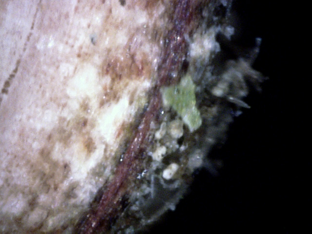
A few things to note here:
1) The most heavily infected phloem area appears to be surrounded by brown, diseased tissue.
2) There is no red cortex tissue here.
3) It looks like an old fragment of bark has old white spores under it (yellow
arrows).
Examination of the bark of a twig can either be helpful or a waste of time.
Often, the crucial factor is knowing exactly where on the bark to look.
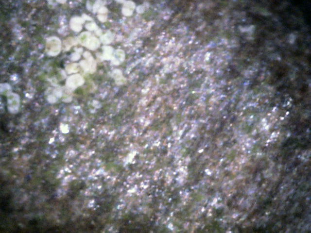
In the photos below, a random location was selected along with an area that past
experience has show to be reliable as hosting numerous white canker spores.
 ←
←
9
Much of the twig surface had a rough glassy look.
But spore patches were also sometimes present (red arrow), as shown in this patch of bark.
10
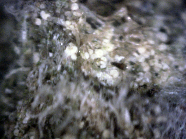
Another patch of spores.
It appears as if these spores had landed and then sent out hyphae to infect the tree through its bark.
11
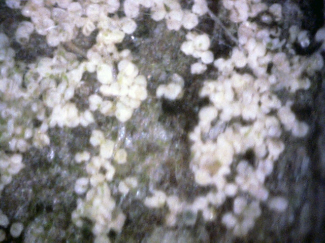
This large patch of spores was located about a foot from the branch end, at a branch junction,
which is the usual area for large bodies of spores to be found.
These spores appear to be old.
In summary, while this eastern red cedar tree has a strong white canker infection,
it is very difficult to determine this by a visual examination of the tree.
Even though the canker is present within the leaves, it cannot be seen visually or
with a microscopic surface inspection since it is buried deeply within the leaf.
As in other infected conifers, leaf and twig cross-sections show diffuse patches
of white canker infection. Twig cross-sections show white canker growths within the outer
phloem, the cortex, and within the outer bark. It's the phloem disrupution that
chokes off nutrients to the tree.
But really strong evidence of white canker infection comes from a 400x microscopic inspection of key areas of the branch's bark. Here we see many areas having a high population of white canker spores.
But really strong evidence of white canker infection comes from a 400x microscopic inspection of key areas of the branch's bark. Here we see many areas having a high population of white canker spores.